Skeleton Bones Labeled Back : Diagram Of The Human Skeletal System Infographic Live Science / The most common variations include sutural (wormian) bones, which are located along the sutural lines on the back of the skull, and sesamoid bones which develop within some tendons, mainly in the hands and feet.
Skeleton Bones Labeled Back : Diagram Of The Human Skeletal System Infographic Live Science / The most common variations include sutural (wormian) bones, which are located along the sutural lines on the back of the skull, and sesamoid bones which develop within some tendons, mainly in the hands and feet.. Almost every skeletal muscle works by pulling two or more bones either closer together or further apart. When autocomplete results are available use up and down arrows to review and enter to select. The human skeletal system consists of all of the bones, cartilage, tendons, and ligaments in the body. In this post, we will discuss the cranial bones and sutures along with their anatomy and landmarks using labeled diagrams. The remaining 7 bones in the head (6 auditory ossicles and 1 hyoid bone) do not articulate with the rest of the skull, and they are often referred to as accessory bones of the skull as a result.
The bones of the appendicular skeleton provide support and flexibility at the joints and anchor the muscles that move the limbs. By tightening and relaxing, the skeletal muscles create movement. Facet joints connect each vertebra, with fluid supporting. The cervical vertebrae, the thoracic vertebrae, the lumbar vertebrae, the sacrum and the coccyx.these sections total 33 vertebrae which function together to aid locomotion and posture as well as providing support and protection. The heart muscle, smooth muscles, and skeletal muscles.
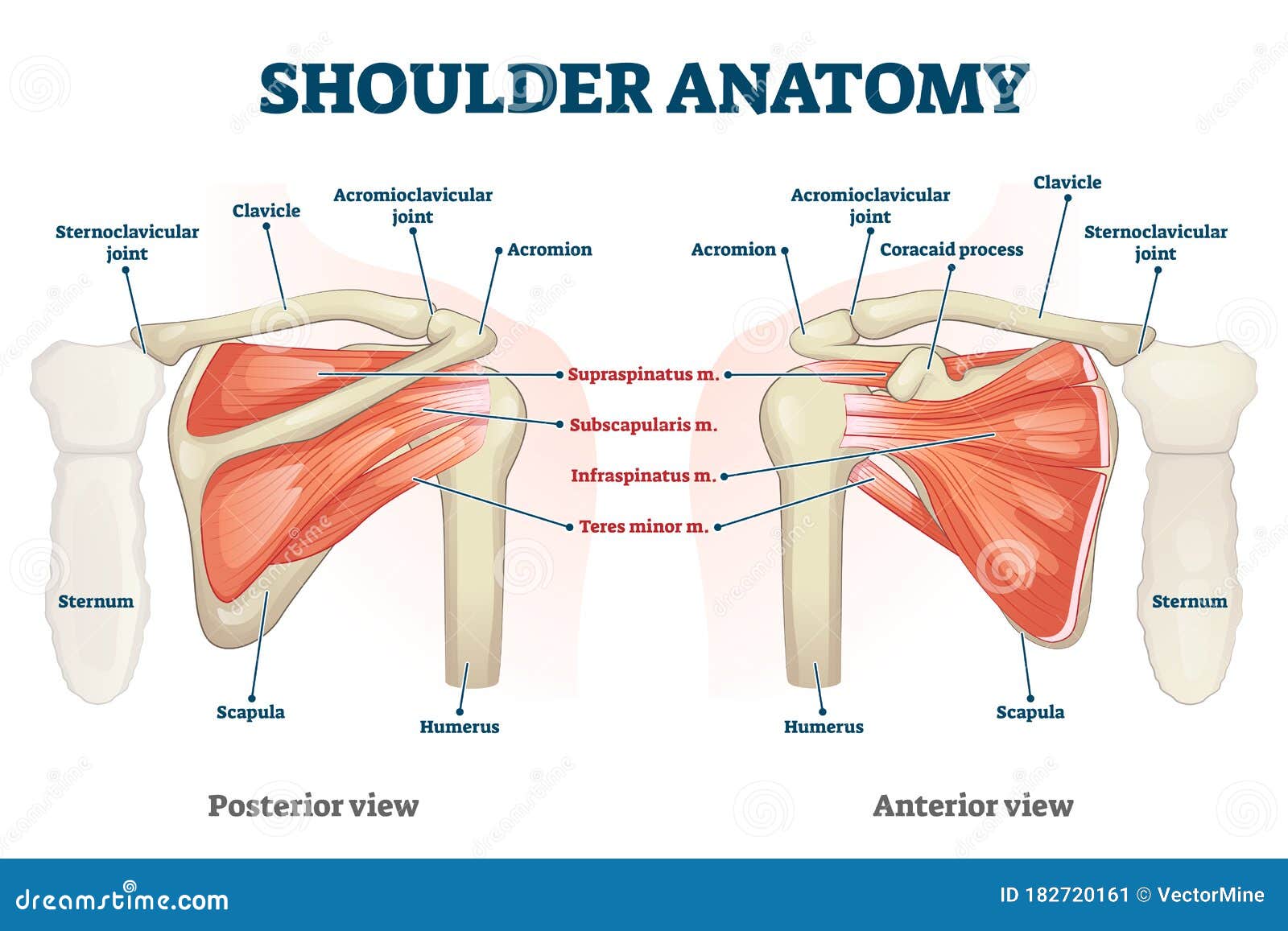
Shoulder Anatomy Vector Illustration Labeled Skeleton And Muscle Scheme Stock Vector Illustration Of Body Back 182720161 from thumbs.dreamstime.com Using this atlas of human anatomy of the spine and back. The bones of the skeletal system act as attachment points for the skeletal muscles of the body. The spine's four sections, from top to bottom, are the cervical (neck), thoracic (abdomen,) lumbar (lower back), and sacral (toward tailbone). Photos of the skeleton bones diagram anatomy bones diagram axial skeleton bones bones of the skeleton quiz human body bones diagram labeled diagram skeleton skeleton diagram with bone names skeleton system bones skull bones diagram. Throughout the spine, intervertebral discs made of. Make you have what you looking for. The most common variations include sutural (wormian) bones, which are located along the sutural lines on the back of the skull, and sesamoid bones which develop within some tendons, mainly in the hands and feet. Some individuals may also have additional (i.e., supernumerary) cervical ribs or lumbar vertebrae.
An opening in the anterior surface of the sacrum
Diagram of a human female skeleton, back view. Altogether, the skeleton makes up about 20 percent of a person's body weight. Throughout the spine, intervertebral discs made of. These bones are connected at the back with specialized joints. Human body anatomy female female anatomy muscle shoulder blade pain anatomy back muscles bones man female anatomy body muscles in a body female anatomy muscole shoulder concept muscular sysyem. Touch device users, explore by touch or with swipe gestures. Numerous muscles, ligaments and tendons support the spine, providing it with flexibility and a great range of motion. An opening in the anterior surface of the sacrum The vertebral column is the defining characteristic of a vertebrate in which the notochord (a flexible rod of uniform composition) found in all chordates has been replaced by a segmented series of bone: The sacrum and two os coxae bones form the pelvis: The cervical vertebrae, the thoracic vertebrae, the lumbar vertebrae, the sacrum and the coccyx.these sections total 33 vertebrae which function together to aid locomotion and posture as well as providing support and protection. Vertebrae, bones, joints, ligaments, muscles, muscular system, fascia, arteries, veins, nerves and various adjacent organs. The most common variations include sutural (wormian) bones, which are located along the sutural lines on the back of the skull, and sesamoid bones which develop within some tendons, mainly in the hands and feet.
While many of us take the benefits of a healthy spine for granted, spinal pain is a sharp reminder of how much we depend on our back in daily life. Vertebrae, bones, joints, ligaments, muscles, muscular system, fascia, arteries, veins, nerves and various adjacent organs. It is formed by 5 fused vertebrae; The vertebral column is the defining characteristic of a vertebrate in which the notochord (a flexible rod of uniform composition) found in all chordates has been replaced by a segmented series of bone: Using this atlas of human anatomy of the spine and back.
File Human Skeleton Front En Svg Wikipedia from upload.wikimedia.org The vertebral column of the lower back includes the five lumbar vertebrae, the sacrum, and the coccyx. The human skeleton, like that of other vertebrates, consists of two principal subdivisions, each with origins distinct from the others and each presenting certain individual features.these are (1) the axial, comprising the vertebral column—the spine—and much of the skull, and (2) the appendicular, to which the pelvic (hip) and pectoral (shoulder) girdles and the bones and cartilages of the. Some individuals may also have additional (i.e., supernumerary) cervical ribs or lumbar vertebrae. Maxilla (2) zygomatic (2) mandible (1) Five bones in the lower back—the lumbar spine the spinal column combines strong bones, unique joints, flexible ligaments and tendons, large muscles and highly sensitive nerves. In this post, we will discuss the cranial bones and sutures along with their anatomy and landmarks using labeled diagrams. Beside that, we also come with more related ideas such labeled human skeleton diagram back, skull bones worksheet and skeletal system matching worksheet. Human body anatomy female female anatomy muscle shoulder blade pain anatomy back muscles bones man female anatomy body muscles in a body female anatomy muscole shoulder concept muscular sysyem.
On anatomical parts the user can choose to display the various structures in colored illustrations of the anatomy of the back and spine:
The spine is composed of 33 bones called vertebrae, which stack together to form the spinal canal. Throughout the spine, intervertebral discs made of. There are three different types of muscles in the body: The bones of the back, together, make up the vertebral column.the vertebral column is made up of 5 sections: These bones are connected at the back with specialized joints. The heart muscle, smooth muscles, and skeletal muscles. The vertebral column (spine) is the bony core of the back. This protects the spinal cord inside. Altogether, the skeleton makes up about 20 percent of a person's body weight. Vertebrae separated by intervertebral discs. They support bones, in this case, the vertebrae. The vertebral column is the defining characteristic of a vertebrate in which the notochord (a flexible rod of uniform composition) found in all chordates has been replaced by a segmented series of bone: Whilst each section of the vertebral column consists of.
The remaining 7 bones in the head (6 auditory ossicles and 1 hyoid bone) do not articulate with the rest of the skull, and they are often referred to as accessory bones of the skull as a result. The red lines point individual bones and the names are writen in singular, the blue lines conect to group of bones and are in plural form. Vertebrae, bones, joints, ligaments, muscles, muscular system, fascia, arteries, veins, nerves and various adjacent organs. Make you have what you looking for. They support bones, in this case, the vertebrae.
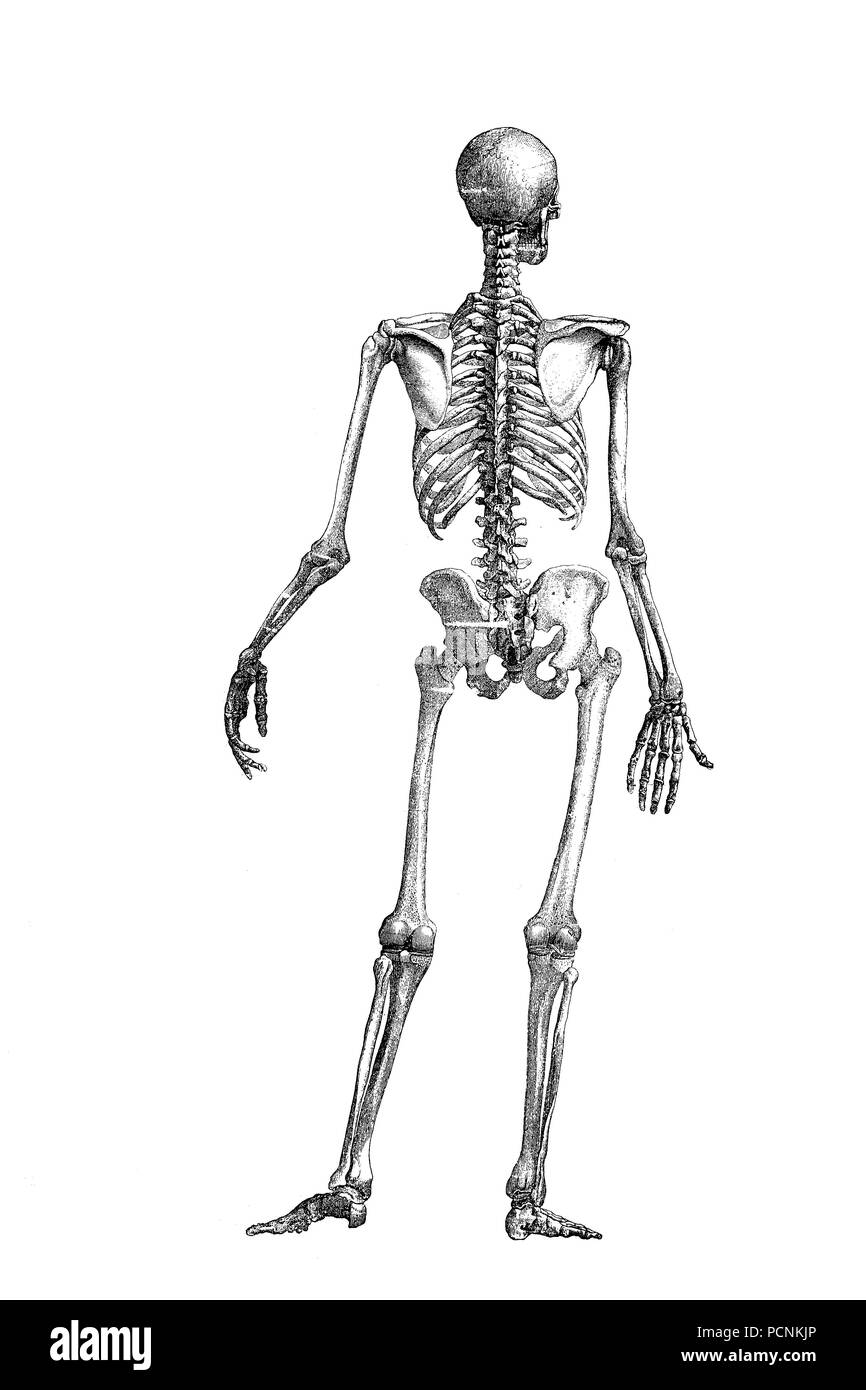
Human Skeleton Back High Resolution Stock Photography And Images Alamy from c8.alamy.com The most common variations include sutural (wormian) bones, which are located along the sutural lines on the back of the skull, and sesamoid bones which develop within some tendons, mainly in the hands and feet. They support bones, in this case, the vertebrae. Almost every skeletal muscle works by pulling two or more bones either closer together or further apart. Axial skeleton (80 bones) skull (28) cranial bones. The heart muscle, smooth muscles, and skeletal muscles. There are three different types of muscles in the body: An opening in the anterior surface of the sacrum A triangular bone that is the posterior skeletal element forming the pelvis:
Vertebrae separated by intervertebral discs.
Vertebrae, bones, joints, ligaments, muscles, muscular system, fascia, arteries, veins, nerves and various adjacent organs. This bone is shaped like a triangle that fits between the two halves of the pelvis, connecting the spine to the lower half of the body. The anatomy of the back refers to the muscles of the back, as well as the bones of the scapulae, ribcage, and spine.covering an expanse from the neck to the tailbone, the back muscles are responsible for a broad range of functions, from extending the spine to shrugging the. The posterior (or back) aspect of the body, and medial (or inside) aspects of the pedicle, and the anterior (or front) lamina form a protective bony ring, called the spinal canal, around the very important dural sac. Maxilla (2) zygomatic (2) mandible (1) The human skeletal system consists of all of the bones, cartilage, tendons, and ligaments in the body. Almost every skeletal muscle works by pulling two or more bones either closer together or further apart. This protects the spinal cord inside. The sacrum and two os coxae bones form the pelvis: These bones work together to provide flexibility to the trunk, support the muscles of the trunk, and protect the spinal cord and spinal nerves of the back. They support bones, in this case, the vertebrae. Human body anatomy female female anatomy muscle shoulder blade pain anatomy back muscles bones man female anatomy body muscles in a body female anatomy muscole shoulder concept muscular sysyem. It is formed by a chain of 33 interconnected vertebrae and their intervening joints.
Almost every skeletal muscle works by pulling two or more bones either closer together or further apart back bones labeled. The bones of the appendicular skeleton provide support and flexibility at the joints and anchor the muscles that move the limbs.


0 Komentar